The second that can save your life: how CT angiography is changing the way we detect heart disease
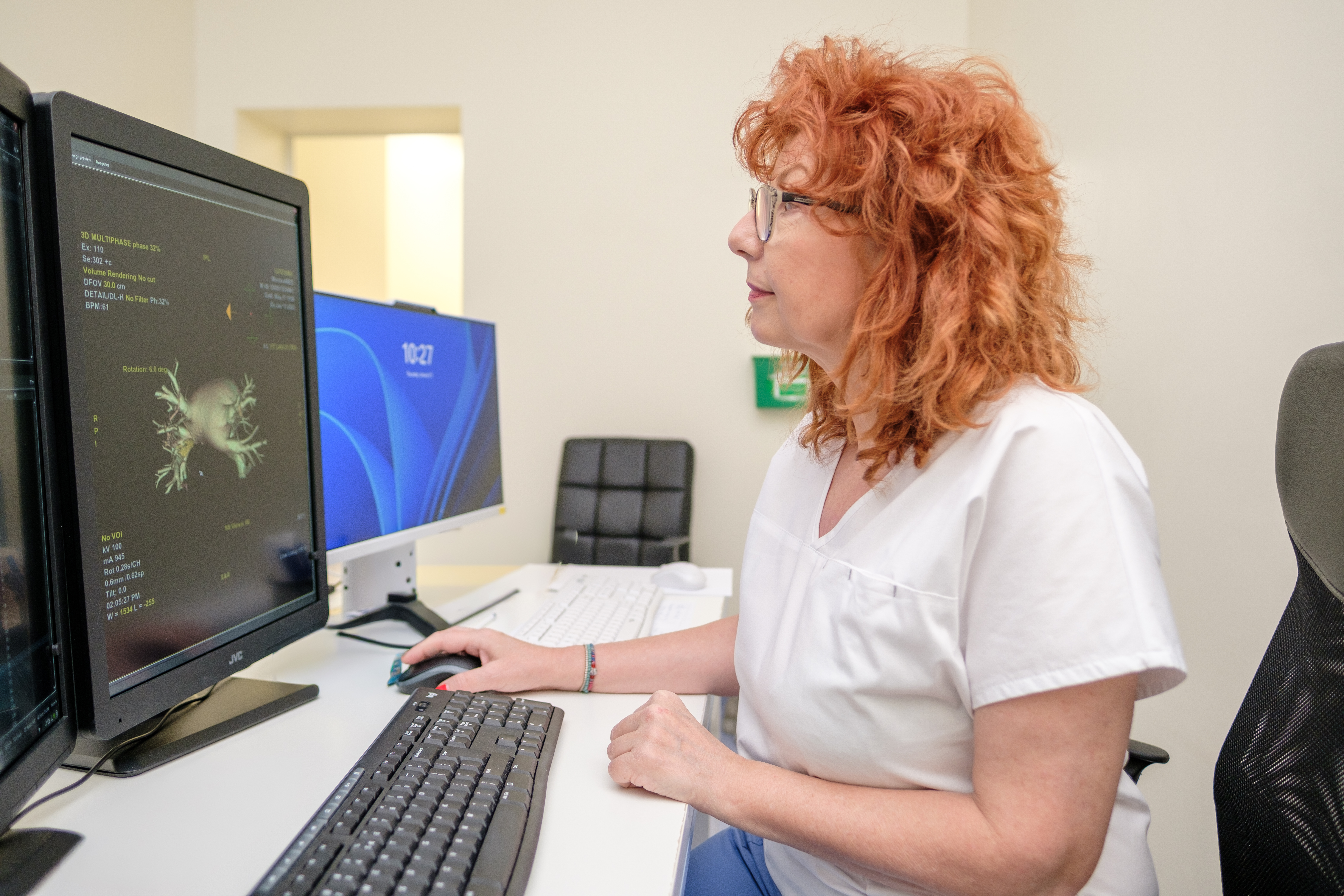
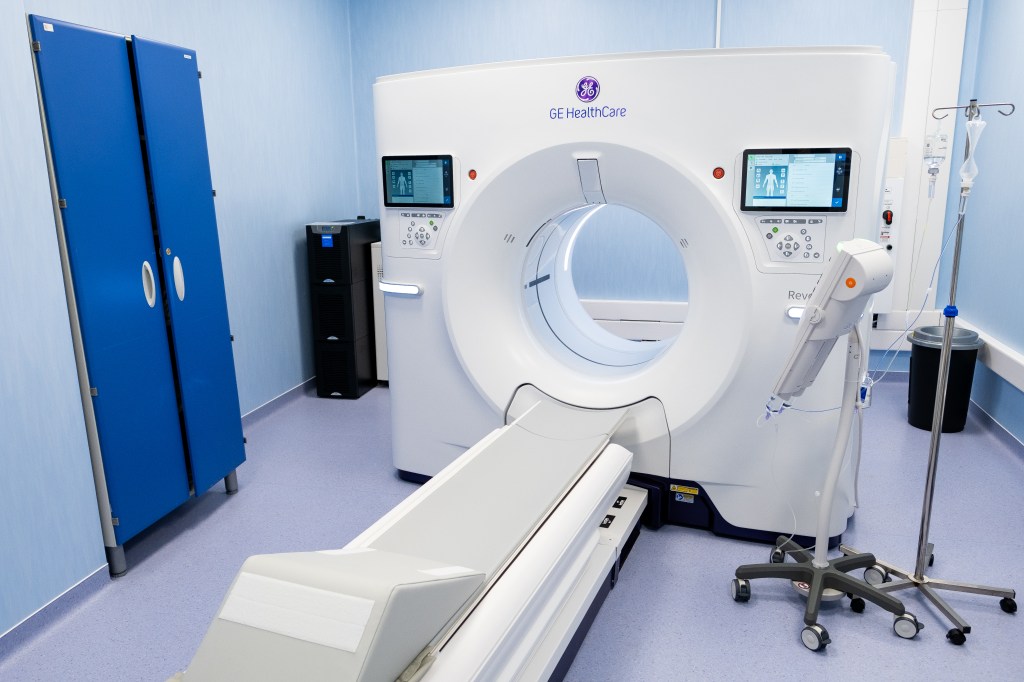
A single second can make the difference between an illness discovered in time and a medical emergency. That's how long a heartbeat lasts, and this interval is enough for the Revolution Vibe CT 512-slice technology, available at MONZA ARES, to capture the full image of the heart. The speed of the scan diminishes the effects of the heart's natural motion, so the images obtained are sharper and more accurate, allowing doctors to assess both heart attack and stroke risk in a single appointment. In a context where cardiovascular diseases can develop for years without symptoms, this rapid diagnostic capability becomes essential.
In many cases, the heart does not announce that it is in danger. You can feel good for years on end. You take your child to school, you go to work, you have your routine tests and everything seems fine. But the paradox is painful: in many cases, cardiovascular diseases they don't always have a clear warning or pain that persists. And sometimes, the first sign of the disease can be even the most serious.
“Cardiovascular diseases can develop slowly and without clinical manifestations until the moment when the situation becomes complicated. Myocardial infarction or stroke can be the first symptom of the disease“, he explains Dr. Ofelia NitaPrimary Physician Radiology and Medical Imaging MONZA ARESwith over 25 years of experience in imaging the cardiovascular system.

A disease that evolves in silence
At the root of many of these events is atherosclerosis – a chronic inflammatory process characterized by the gradual deposition of fats and cholesterol on the artery walls. Over time, these deposits form atheromatous plaques that can narrow blood vessels and reduce flow to the heart. Evolution is slow and, at first, completely silent. That's why many people don't know that their arteries are already damaged.
“Atherosclerosis is a long-term process that can appear at young ages and evolve over a period of years. Clinical manifestations may actually represent the expression of the complication of the disease,” says the doctor.
When symptoms appear, the disease may already be in an advanced stage. In certain situations, the atheroma plaque not only progressively narrows the vessel, but can also rupture, forming a blood clot that suddenly cuts off circulation. This is how a heart attack can occur, even in people who until then had no obvious reasons for concern.
In this context, early diagnosis becomes more than a recommendation, it becomes a prevention strategy. And one of the investigations that can bring decisive information is the coronary angio-CT, a non-invasive imaging method that allows the direct evaluation of the arteries of the heart.
Why are classic investigations not enough?
Classic cardiological investigations, such as the electrocardiogram, cardiac ultrasound or stress test, show how the heart is working. Coronary CT angiography, however, shows the arteries directly.
“It is the only non-invasive method that can provide a complete picture of the coronary arteries, both the inside of the vessel and the vascular wall. We can identify not only the narrowing, but also the structure of the atheroma plaque. Soft, non-calcified lipid plaques are considered to be at high risk of complications,” explains Dr. Niță.
This difference is essential, because infarction is not exclusively produced by severe stenoses. Sometimes an apparently moderate but unstable plaque may be the trigger for the acute event.
Through angio-CT, the doctor can evaluate the entire coronary bed in a single examination: where the plaques are, how many there are and how stable they are, information that cannot be completely obtained by other non-invasive methods.
In addition, the cervico-cerebral vessels can also be evaluated in the same session, providing a complete picture of both heart attack and stroke risk. This integrated approach allows for a global assessment of cardiovascular risk, in a single appointment and in a very short time. This is an important advantage especially for intermediate risk patients, in whom the disease can evolve for years without symptoms.
The Myths That Make Us Procrastinate – Reality Versus Perception?
Many patients postpone seeing a doctor based on seemingly logical beliefs. The reality, however, is more nuanced. Studies of asymptomatic populations have shown that a significant percentage of people without symptoms may already have subclinical atherosclerosis – changes that do not yet produce visible manifestations. And the moment it becomes symptomatic can sometimes be brutal.
The most common belief that delays diagnosis is simple: if it doesn't hurt, it's nothing. Or its version: I'm active, I feel good, I have nothing to worry about. “In reality, the stenosis phase is already an advanced stage of atherosclerotic disease. The intention is to detect the disease early, precisely to be able to intervene before complications”, explains Dr. Niță.
Beyond these beliefs there is another fear that causes patients to procrastinate: fear of investigations – radiation or contrast material. It's a natural reaction. But, currently, the investigation is carried out with state-of-the-art equipment, and the level of safety is high.
“The examination does not involve a high level of radiation exposure and no greater risks related to the contrast material than in other CT investigations. In addition, we noted a reduction in the amount of contrast material by 30-40% compared to conventional equipment. There are algorithms and filters that allow us to very carefully control the exposure.” explains Dr. Ofelia Niță.
In patients with a history of allergies, the investigation can be performed safely, with allergy evaluation and, if necessary, preventive treatment before the procedure, so that the investigation is carried out safely.
Why it matters to know ahead of time – even a second can make a difference
In most cases, the benefit of finding out the condition of the arteries of the heart in time far outweighs the fears of the investigation. Perhaps the most surprising detail is the speed of the examination. At MONZA ARES, the investigation is carried out with Revolution Vibe CT 512-slice – the first system of this type in the private environment in Romania, capable of capturing the image of the heart in a single beat.
“The heart scan takes, on average, one second. That's how long it takes for the machine to capture the image of the heart. Including patient preparation, the whole procedure takes about 10 minutes. It's a paradigm shift in the way we approach investigations”says the doctor.
Cardiovascular disease does not start on the day of the heart attack, it develops years before. Angio-CT is especially recommended for people with risk factors such as hypertension, diabetes, high cholesterol, smoking or family history. It is considered a first-line method, especially in intermediate or low-risk patients – people who, in the absence of an imaging evaluation, could go with this type of problem unnoticed for years.
Angio-CT does not replace a healthy lifestyle or cardiological consultation, but it can complement the traditional assessment, providing information that cannot be obtained otherwise. The difference between a problem discovered in time and a medical emergency can lie in a decision made at the right time – for you and those who depend on you – to investigate before the disease manifests itself. And sometimes even a second can count.
Article supported by MONZA ARES