[P] What the naked eye can't see can matter: how the dental operating microscope is changing dental treatments

Precision has become essential in modern dentistry to achieve sustainable and predictable results. The dental operating microscope, initially used almost exclusively in root canal treatments, is starting to be used more and more frequently in other stages of dental treatments – from consultation and diagnosis, to restorations and dental aesthetics.
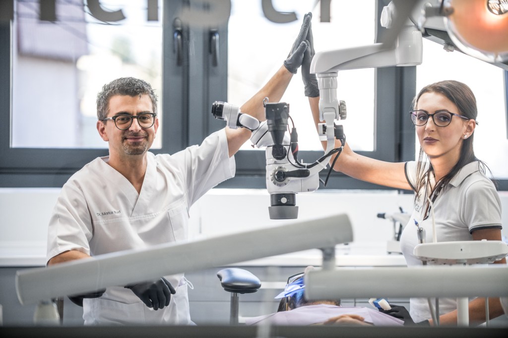
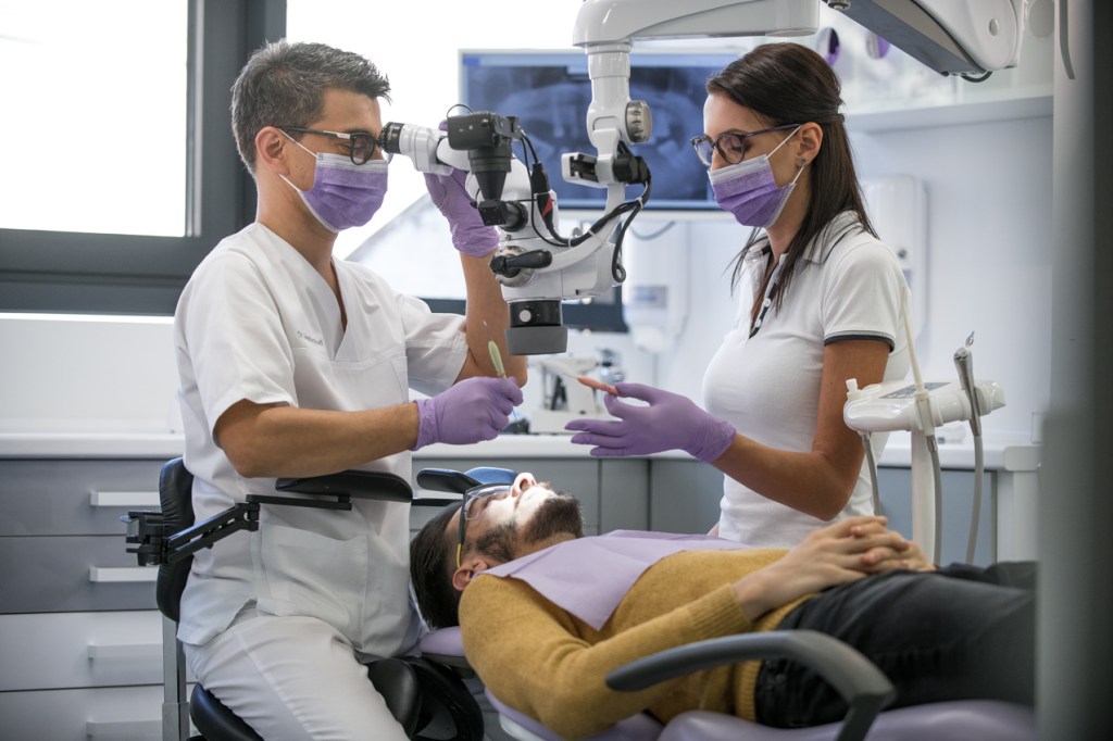
“The dental operating microscope is not just a tool for endodontics. It fundamentally changes the way we see and treat the tooth, regardless of the procedure. The image can be magnified up to 20 times, and the lighting is incomparably better than with the naked eye. Exactly what a dentist needs to pay attention to details: to see bigger and clearer”, explains Marius Bud, dentist, founder of the MB Dental clinic and head of works at the University of Medicine and Pharmacy “Iuliu Hatieganu” from Cluj-Napoca.
Digital planning and the dental microscope: more precise and less invasive aesthetic dental treatments
One of the first stages where the microscope brings benefits is the dental consultation. Optical magnification and direct illumination allow the identification of details that are hard to see with the naked eye: microcracks, early caries, fine filling defects or infiltration areas.
Studies show that the use of optical magnification improves diagnostic accuracy and the ability to detect early dental lesions, which allows for more conservative and better targeted interventions.
Minimally invasive removal of dental caries
In the treatment of caries, working under the microscope allows a clear demarcation between healthy and damaged tooth tissue. Thus, the doctor can remove the caries completely, but with minimal loss of tooth substance.
“Working under magnification, you can control every movement. You're strictly removing what's affected, without unnecessarily weakening the tooth structure,” says Dr. Bud.
Research shows that working under the microscope increases the precision of clinical procedures and reduces the risk of leaving damaged tissue compared to classical techniques.
Tighter and more durable seals
Performing obturations (seals) under a microscope allows for much better control of material adaptation, elimination of air bubbles and correct edge finishing. These details directly influence the tightness and lifespan of the restoration.
Data from the specialized literature indicate a significant reduction in marginal defects and an increase in the longevity of fillings made under optical magnification.
Preparing teeth for crowns and other prosthetic restorations
In dental prosthetics, the microscope helps in a very precise preparation of teeth for crowns or partial restorations. The edges are clearly defined and the fit of the final work is more predictable.
“A correct preparation of the tooth means a better adapted crown and a lower risk of long-term biological complications,” explains Dr. Marius Bud.
Ceramic veneers and digitally guided dental aesthetics
In dental aesthetics, the microscope naturally integrates with digital smile planning. Software such as Digital Smile Design, including those assisted by artificial intelligence, allow the initial situation to be superimposed with the desired result, using photos, scans and digital simulations.
“Digital planning shows us where we want to go. The microscope helps us prepare the teeth exactly according to the plan, as conservatively as possible, without unnecessary polishing of the enamel,” emphasizes Dr. Marius Bud, from the MB Dental clinic.
This combination of digital planning and microscopic work allows minimally invasive preparations for ceramic veneers, following the established plan and reducing the risk of excessive tooth grinding.
Why working under a microscope is more accurate
Optical magnification not only improves vision but also movement control. Studies show that working under a microscope can increase the accuracy of clinical gestures by up to 40–60% compared to working without magnification, due to superior visual feedback and better control of the work area.
The direct lighting and ergonomic position also help to reduce doctor fatigue and achieve more consistent results.
The benefits for the patient
For the patient, using the microscope means more precise, less invasive and safer treatments. Correctly fitted restorations reduce the risk of infiltration, post-treatment sensitivity and long-term complications.
“Technology does not replace the doctor's experience, but supports it. The microscope helps us to do things more accurately, more controlled and safer,” concludes Dr. Bud.
Clinical experience and integrated approach
MB Dental, a clinic present in Cluj-Napoca and Târgu Mureș, is among the centers that early integrated the dental operating microscope, initially in endodontics, since 2011, and later also in dental restorations, prosthetics and dental aesthetics for veneers.
In addition to clinical work, the team is involved in university education and the continuing education of dentists and promotes the responsible use of modern technology through postgraduate courses.
For patients, choosing a clinic that uses the dental operating microscope and digital smile planning means access to treatments aligned with the current standards of modern dentistry and real care for the preservation of natural teeth.