Chinese researchers have captured the first high-resolution images of human embryos in their first five days of development, a breakthrough that has led to an important discovery about why so many embryos fail to develop in in vitro fertilization clinics, Xinhua reported on Monday, cited by Agerpres.
The study, led by researchers at Tsinghua University in Beijing, was published in the scientific journal Cell.
For couples undergoing in vitro fertilization (IVF) treatment, the road to having a child is not always crowned with success. More than half of fertilized human eggs stop developing before they reach the blastocyst stage, when they are ready to implant in the mother's uterus. This high failure rate has long puzzled scientists.
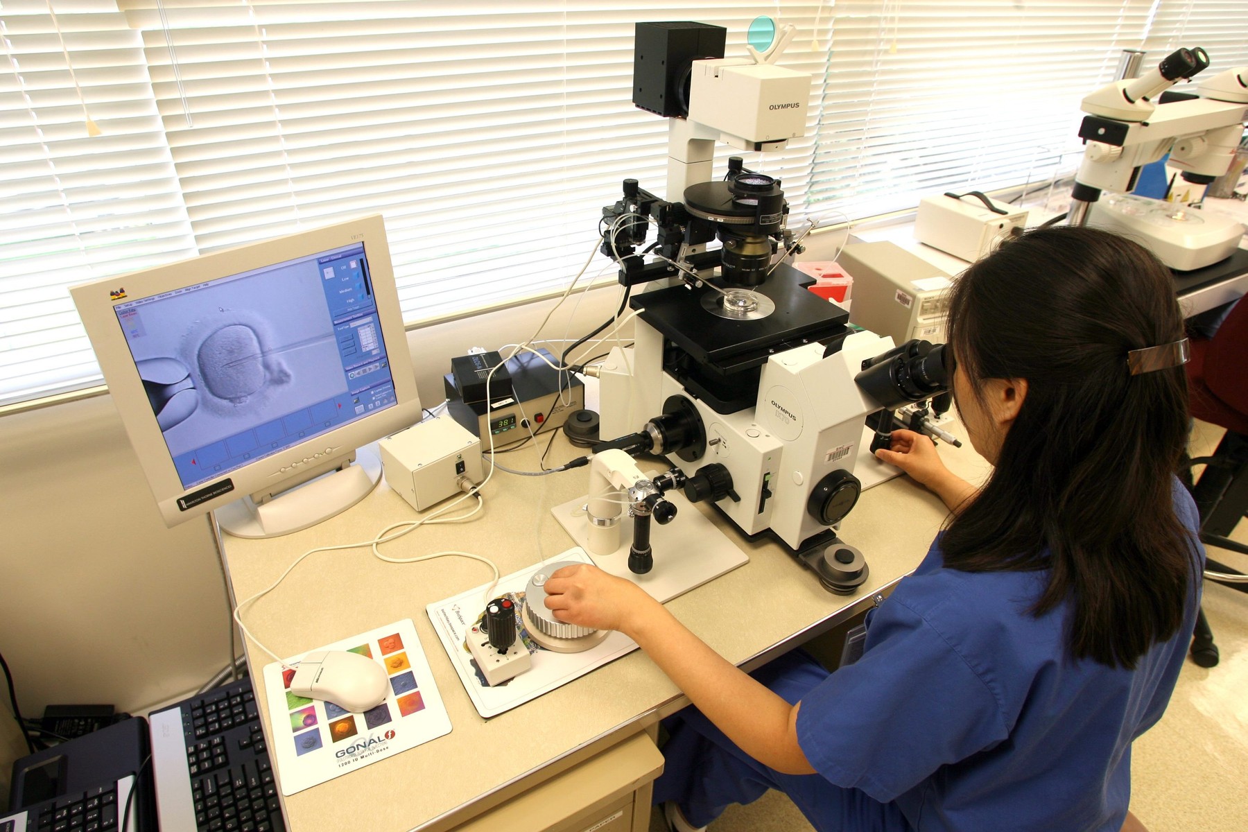
To unravel this mystery, researchers from the Chinese team built a new type of microscope: a double-viewing light beam microscope. This advanced tool can capture clear images of living embryos over a longer period of time without harming them.
Scientists have studied embryos that have stopped developing prematurely
Studying more than 2,000 cell divisions in more than 150 human and monkey embryos, researchers in China found that more than 70 percent of embryos that stopped developing prematurely had problems during the second cell division.
Specifically, a structure called a “spindle,” which looks like a spinning wheel and helps divide genetic material equally, was abnormal in such cases. This caused errors in chromosome segregation, leading to cell cycle arrest within the next few cell divisions.
A second discovery was related to centrosomes, cell organelles that help form the “spindle” and play a key role in those errors. When the number of centrosomes was incorrect, the “spindle” could no longer function properly.
The researchers say the discovery has applications for fertility clinics
Starting from this discovery, Chinese researchers tried to correct the errors that occurred during the second cell division. They administered a small dose of a drug that acts on the replication of centrosomes.
The result was impressive: the percentage of embryos with normal centrosomes increased from 40% to 80%. It is also worth noting that the drug used had no effect on embryos that already had normal centrosomes.
“In the future, this discovery could be used to help prevent early arrest of embryo development in IVF clinics,” said Chun So, an assistant professor at Tsinghua University.
He added that his team will continue to develop advanced imaging technologies to study post-implantation development in humans, which should ultimately lead to new strategies to improve the chances of a successful pregnancy.